Home
/ Rib Cage Anatomy - Three Dimensional View Of Female Rib Cage And Skeletal System Stock Photo Alamy : Bifid ribs occur in up to 8.4% of samoans.
Rib Cage Anatomy - Three Dimensional View Of Female Rib Cage And Skeletal System Stock Photo Alamy : Bifid ribs occur in up to 8.4% of samoans.
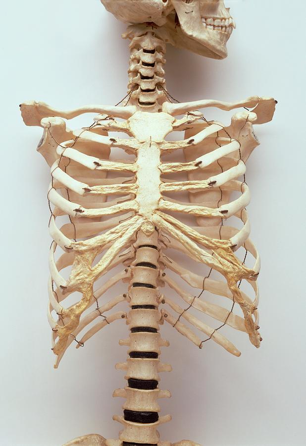
Rib Cage Anatomy - Three Dimensional View Of Female Rib Cage And Skeletal System Stock Photo Alamy : Bifid ribs occur in up to 8.4% of samoans.. The rib cage is a bony structure found in the chest (thoracic cavity). An inhalation is accomplished when the muscular diaphragm, at the floor of the thoracic cavity, contracts and flattens, while the contraction of intercostal muscles lift the rib cage up and out. The costocorporeal joint is where the rib head connects with two adjacent vertebral bodies and the disc between them. Each pair is numbered based on their attachment to the sternum, a bony process at the front of the rib cage which serves as an anchor point. It has a roughened area on its upper surface, from which the serratus anterior muscle originates.
A cervical rib forms from the overdevelopment of the transverse process of a cervical vertebra, typically from the seventh cervical vertebra in the neck known as c7. It is made up of 12 pairs of ribs. Feb 10, 2020 · anatomy. Oct 20, 2020 · rib 2 is thinner and longer than rib 1, and has two articular facets on the head as normal. It encloses the thoracic cavity, which contains the lungs.
Rib Cage Anatomy Function Britannica from cdn.britannica.com The costocorporeal joint is where the rib head connects with two adjacent vertebral bodies and the disc between them. The rib below that is rib 2, and it connects to the t2 thoracic vertebra, and so on. It has a roughened area on its upper surface, from which the serratus anterior muscle originates. See facet joint anatomy animation. Jun 10, 2021 · the thoracic cage (rib cage) is the skeleton of the thoracic wall. An inhalation is accomplished when the muscular diaphragm, at the floor of the thoracic cavity, contracts and flattens, while the contraction of intercostal muscles lift the rib cage up and out. Oct 20, 2020 · rib 2 is thinner and longer than rib 1, and has two articular facets on the head as normal. The sternal end of the rib is cleaved into two.
Oct 20, 2020 · rib 2 is thinner and longer than rib 1, and has two articular facets on the head as normal.
The sternal end of the rib is cleaved into two. An inhalation is accomplished when the muscular diaphragm, at the floor of the thoracic cavity, contracts and flattens, while the contraction of intercostal muscles lift the rib cage up and out. The human rib cage is a component of the human respiratory system. A bifid rib is a congenital abnormality of the rib cage and associated muscles and nerves which occurs in about 1.2% of humans. It has a roughened area on its upper surface, from which the serratus anterior muscle originates. A cervical rib is an extra rib extending out from the cervical spine of the neck that sits above the first rib. The rib below that is rib 2, and it connects to the t2 thoracic vertebra, and so on. Lumbar (or 13th) ribs are a rare anatomical variant and represent transitional vertebrae at the thoracolumbar junction with a prevalence of ~1% 1. This article will look at the osteology of the thoracic vertebrae, examining their characteristic features, joints and clinical correlations. Ten of the twelve ribs connect to strips of hyaline cartilage on the anterior side of the body. Sep 05, 2019 · along with the sternum and ribs, the thoracic spine forms part of the thoracic cage. It is supported by the vertical sternum or. Oct 20, 2020 · rib 2 is thinner and longer than rib 1, and has two articular facets on the head as normal.
The rib below that is rib 2, and it connects to the t2 thoracic vertebra, and so on. It has a roughened area on its upper surface, from which the serratus anterior muscle originates. The costocorporeal joint is where the rib head connects with two adjacent vertebral bodies and the disc between them. A cervical rib forms from the overdevelopment of the transverse process of a cervical vertebra, typically from the seventh cervical vertebra in the neck known as c7. It is supported by the vertical sternum or.
Human Rib Cage Stock Photo Download Image Now Istock from media.istockphoto.com The costocorporeal joint is where the rib head connects with two adjacent vertebral bodies and the disc between them. A cervical rib forms from the overdevelopment of the transverse process of a cervical vertebra, typically from the seventh cervical vertebra in the neck known as c7. Oct 20, 2020 · rib 2 is thinner and longer than rib 1, and has two articular facets on the head as normal. See facet joint anatomy animation. Dec 21, 2020 · anatomy the rib cage has 12 sets of ribs. It presents as an additional rib coming off t13 or l1 (depending on numbering classification) and m. The sternal end of the rib is cleaved into two. The rib cage is a bony structure found in the chest (thoracic cavity).
Feb 10, 2020 · anatomy.
Bifid ribs occur in up to 8.4% of samoans. There are two types of costovertebral joints: The cartilage strips are called costal cartilage ("costal" is the anatomical adjective that refers to the rib) and connect on their other end to the sternum. It is supported by the vertical sternum or. Oct 20, 2020 · rib 2 is thinner and longer than rib 1, and has two articular facets on the head as normal. The rib below that is rib 2, and it connects to the t2 thoracic vertebra, and so on. This article will look at the osteology of the thoracic vertebrae, examining their characteristic features, joints and clinical correlations. An inhalation is accomplished when the muscular diaphragm, at the floor of the thoracic cavity, contracts and flattens, while the contraction of intercostal muscles lift the rib cage up and out. A cervical rib forms from the overdevelopment of the transverse process of a cervical vertebra, typically from the seventh cervical vertebra in the neck known as c7. Each pair is numbered based on their attachment to the sternum, a bony process at the front of the rib cage which serves as an anchor point. Dec 21, 2020 · anatomy the rib cage has 12 sets of ribs. It presents as an additional rib coming off t13 or l1 (depending on numbering classification) and m. The human rib cage is a component of the human respiratory system.
Ten of the twelve ribs connect to strips of hyaline cartilage on the anterior side of the body. Jun 10, 2021 · the thoracic cage (rib cage) is the skeleton of the thoracic wall. Bifid ribs occur in up to 8.4% of samoans. Each pair is numbered based on their attachment to the sternum, a bony process at the front of the rib cage which serves as an anchor point. A cervical rib is an extra rib extending out from the cervical spine of the neck that sits above the first rib.
Human Rib Cage Photograph By Dorling Kindersley Uig from images.fineartamerica.com It has a roughened area on its upper surface, from which the serratus anterior muscle originates. Each pair is numbered based on their attachment to the sternum, a bony process at the front of the rib cage which serves as an anchor point. There are two types of costovertebral joints: An inhalation is accomplished when the muscular diaphragm, at the floor of the thoracic cavity, contracts and flattens, while the contraction of intercostal muscles lift the rib cage up and out. Jun 10, 2021 · the thoracic cage (rib cage) is the skeleton of the thoracic wall. Oct 20, 2020 · rib 2 is thinner and longer than rib 1, and has two articular facets on the head as normal. It is supported by the vertical sternum or. Dec 21, 2020 · anatomy the rib cage has 12 sets of ribs.
The sternal end of the rib is cleaved into two.
The costocorporeal joint is where the rib head connects with two adjacent vertebral bodies and the disc between them. Bifid ribs occur in up to 8.4% of samoans. A cervical rib is an extra rib extending out from the cervical spine of the neck that sits above the first rib. The sternal end of the rib is cleaved into two. The thoracic cage takes the form of a domed bird cage with the horizontal bars formed by ribs and costal cartilages. Feb 10, 2020 · anatomy. Each pair is numbered based on their attachment to the sternum, a bony process at the front of the rib cage which serves as an anchor point. Dec 21, 2020 · anatomy the rib cage has 12 sets of ribs. It is formed by the 12 thoracic vertebrae, 12 pairs of ribs and associated costal cartilages and the sternum. Oct 20, 2020 · rib 2 is thinner and longer than rib 1, and has two articular facets on the head as normal. The cartilage strips are called costal cartilage ("costal" is the anatomical adjective that refers to the rib) and connect on their other end to the sternum. It is supported by the vertical sternum or. The rib below that is rib 2, and it connects to the t2 thoracic vertebra, and so on.